Patient Presentation: A 23-year-old obese female was diagnosed with idiopathic intracranial hypertension (IIH) and referred to neurosurgery for ventriculoperitoneal shunt. A baseline ocular examination was performed prior to the procedure.
On examination, vision was 20/200 in the right eye, and 20/40 in the left eye. There was a right relative afferent pupillary defect. Slit lamp examination was normal.
A dilated fundus examination was performed demonstrating the following:
.png)
Glaucoma
Case 8
Patient Presentation: A 68-year-old male was referred by his optometrist with concern for glaucoma. Visual acuity was 20/20 OU. IOP measured 15 mmHg OD and 16 mmHg OS, with one glaucoma medication class in each eye. His angles were open bilaterally on gonioscopy.
OCT ONH/RNFL and ganglion cell complex (GCC) were performed and are shown below:
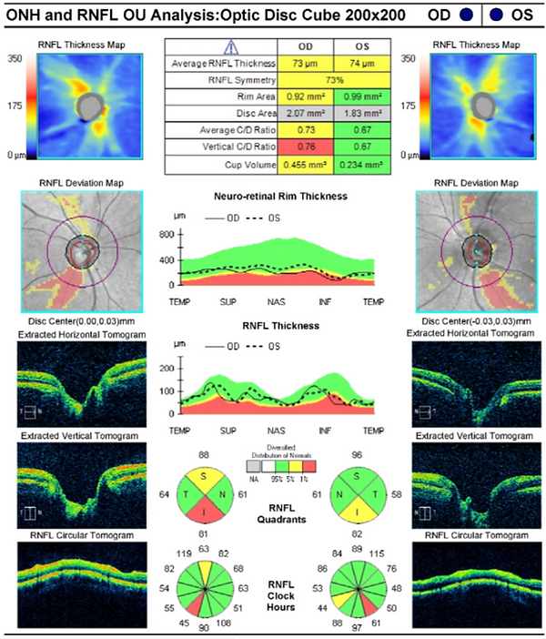

Question 1: What are the most striking abnormalities on OCT ONH/RNFL?
Question 2: What visual field defect would you expect to see in this patient?
Question 3: What would be your next steps if the patient had the OCT RNFL/GCC findings described above and a normal, reliable 24-2 Humphrey visual field tes?
Question 4: Why is it important to use multiple testing modalities (RNFL OCT, GCC OCT, and visual field testing) when assessing glaucoma?
References:
-
Hood DC. Improving our understanding, and detection, of glaucomatous damage: An approach based upon optical coherence tomography (OCT). Prog Retin Eye Res. 2017;57:46-75. doi:10.1016/j.preteyeres.2016.12.002
-
Hood Visual Science Lab. “Module 1: Background Information and the Circumpapillary (cpRNFL) Report.” Jul 21, 2021. Accessed December 5 2025 from: https://hoodvisualscience.psychology.columbia.edu/content/module-1-background-information-and-circumpapillary-cprnfl-report
Learning Objectives:
-
Macular Vulnerability Zone (MVZ): This inferior-temporal zone is highly susceptible in glaucoma; damage here produces paracentral/central field loss and early GCC loss.
-
Multimodal concordance: True glaucoma evaluations require structural-functional correlation (RNFL thinning + ganglion cell loss + matching visual field defect). This is essential for accurate diagnosis and confirmation.
-
Central visual field defects automatically place patients into advanced glaucoma per the Hodapp–Parrish-Anderson classification.
-
HVF 10-2 and 24-2c modules can detect macular damage through additionally tested central points which may be missed on standard 24-2 testing