Patient Presentation: A 23-year-old obese female was diagnosed with idiopathic intracranial hypertension (IIH) and referred to neurosurgery for ventriculoperitoneal shunt. A baseline ocular examination was performed prior to the procedure.
On examination, vision was 20/200 in the right eye, and 20/40 in the left eye. There was a right relative afferent pupillary defect. Slit lamp examination was normal.
A dilated fundus examination was performed demonstrating the following:
.png)
Retina
Case 64
Patient Presentation: A 24-year-old male was referred because of painless blurred vision (OD > OS) for 1 month. His past medical history was significant for a pineal tumour which was treated by craniospinal radiation (36 Gy, 54 Gy and then 59.4 Gy). He did not have diabetes or hypertension. On exam, visual acuity was 20/150 OD and 20/40-1 OS. Colour vision was 0/12 OD and 7/14 OS. There was no RAPD. OCT macula was performed and shown below.
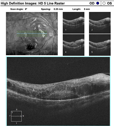

These abnormalities correlated with findings on fundoscopy:


Question: What should immediately be done in the office?
The patient’s blood pressure was 130s/80s.
Question: What is the likely cause of this patient’s presentation?
Question: How should this patient be managed?
Question: If this patient were to undergo further craniospinal radiation, how would you treat to prevent worsening radiation retinopathy?
References:
-
Danesh-Meyer HV. Radiation-induced optic neuropathy. J Clin Neurosci. 2008;15(2):95-100.
-
Fallico M, Chronopoulos A, Schutz JS, Reibaldi M. Treatment of radiation maculopathy and radiation-induced macular edema: A systematic review. Surv Ophthalmol. 2021;66(3):441-460.
-
Giuliari GP, Sadaka A, Hinkle DM, Simpson ER. Current treatments for radiation retinopathy. Acta Oncol. 2011;50(1):6-13.
-
Sahoo NK, Ranjan R, Tyagi M, Agrawal H, Reddy S. Radiation Retinopathy: Detection and Management Strategies. Clin Ophthalmol. 2021;15:3797-3809. Published 2021 Sep 8. doi:10.2147/OPTH.S219268
Learning Objectives:
-
Interpreting OCT macula images in patients with radiation retinopathy
-
Identify patients at risk for radiation retinopathy
-
Understand principles in management for patients with radiation retinopathy