top of page
Patient Presentation: A 23-year-old obese female was diagnosed with idiopathic intracranial hypertension (IIH) and referred to neurosurgery for ventriculoperitoneal shunt. A baseline ocular examination was performed prior to the procedure.
On examination, vision was 20/200 in the right eye, and 20/40 in the left eye. There was a right relative afferent pupillary defect. Slit lamp examination was normal.
A dilated fundus examination was performed demonstrating the following:
.png)
Retina
Case 27
Contributor: Dr. Sultan Aldrees, MD
Patient Presentation: A 41-year-old male presents to clinic with a longstanding history of decreased vision in both eyes. On examination, visual acuity is 20/200 in the right eye and 20/250 in the left eye. There was no RAPD. Intraocular pressures were normal. Slit lamp exam was unremarkable. Fundus exam is shown through the Optos® photos. FAF and OCT were also ordered, shown below:


Question: Which of the following features are seen in the above OCT scan?
Question: In the FAF, there are areas of hyper and hypo-autofluorescence. What is the significance of these findings?
Question: Given the patient’s young age and longstanding history of poor vision, you perform genetic testing that is positive for a mutation in ABCA4 gene. Given this, what is the likely diagnosis?
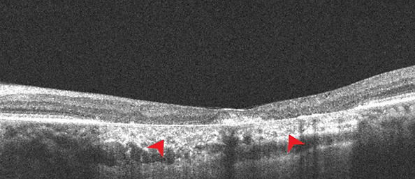
Question: What is the red arrow pointing to in the OCT above?

Question: What layer of the retina is the yellow arrow pointing to?
Question: SD can lead to bull’s eye atrophic maculopathy. Can you list four other differential diagnosis of bull’s eye maculopathy?

Question: A fluorescein angiogram (FA) was obtained for this patient, shown below. What is the classic FA sign seen in Stargardt disease?
Learning Objectives:
1. To be able to recognize the OCT, FAF and FA features of Stargardt disease. Understanding the pathophysiology of the disease will help in remembering these features.
bottom of page