Patient Presentation: A 23-year-old obese female was diagnosed with idiopathic intracranial hypertension (IIH) and referred to neurosurgery for ventriculoperitoneal shunt. A baseline ocular examination was performed prior to the procedure.
On examination, vision was 20/200 in the right eye, and 20/40 in the left eye. There was a right relative afferent pupillary defect. Slit lamp examination was normal.
A dilated fundus examination was performed demonstrating the following:
.png)
Neuro-Ophthalmology
Case 20
Contributor: Korolos Sawires
Patient Presentation: A 54-year-old woman presented to ophthalmology for a routine eye examination. The patient denied ocular complaints or changes over the past year. The best-corrected distance visual acuity was 20/20. Fundus examination was performed, which revealed the following:


Question: Please describe the above fundus photographs. Are there any notable abnormalities? What would be a logical next step?
SS-OCT’s over the macula and optic were performed in the right and left eye, and are shown below:

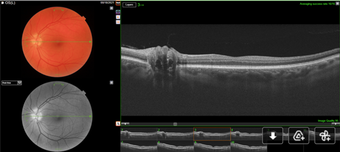
Question: Based on the above SS-OCT scans, what abnormality can be seen?
Question: Is this a case of pseudo-papilledema or papilledema?
Question: What is the diagnosis?
References:
-
Fraser JA, Bursztyn LLC. Optical coherence tomography in optic disc drusen. Ann Eye Sci 2020;5:5.
-
Malmqvist L, Bursztyn L, Costello F, Digre K, Fraser JA, Fraser C, Katz B, Lawlor M, Petzold A, Sibony P, Warner J, Wegener M, Wong S, Hamann S. The Optic Disc Drusen Studies Consortium Recommendations for Diagnosis of Optic Disc Drusen Using Optical Coherence Tomography. J Neuroophthalmol. 2018 Sep;38(3):299-307. doi: 10.1097/WNO.0000000000000585. PMID: 29095768.
-
Rebolleda G, Kawasaki A, de Juan V, Oblanca N, Muñoz-Negrete FJ. Optical Coherence Tomography to Differentiate Papilledema from Pseudopapilledema. Curr Neurol Neurosci Rep. 2017 Aug 17;17(10):74. doi: 10.1007/s11910-017-0790-6. PMID: 28819712.
-
Wester ST, Fantes FE, Lam BL, Anderson DR, McSoley JJ, Knighton RW. Characteristics of optic nerve head drusen on optical coherence tomography images. Ophthalmic Surg Lasers Imaging. 2010 Jan-Feb;41(1):83-90. doi: 10.3928/15428877-20091230-15. PMID: 20128575.
Learning Objectives:
-
To identify OCT features of optic disc drusen and differentiate between ODD mimics
-
To differentiate optic disc drusen from papilledema on OCT